BIO 102 MENU
syllabus 
1 - origin
2 - biomol.
3 - biomol2
4 - biomol3
5 - viruses
6 - prokaryon
7 - endosym
8 - eukaryon
9 - energy
10 - mitosis
11 - meiosis
12 - reprod
13 - genetics
14 - humgene
15 - humge2
16 - evolution
17 - evolutio2
18 - diversity
19 - diversi2
20 - tissues
21 -digestive
22 - respirat
23 - circul
24 - excret
25 - endocr
26 - receptors
27 - nervsys
Quizzes
Bio 103 Lab
(full title of lecture appears in status bar on the top or at the bottom of your window)

Biology 102 - General Biology
Eukaryotic Cells
At this time five kingdoms are recognized. The most primitive organisms are in the Kingdom Monera, made up exclusively of prokaryotes. Protista, Fungi, Plants, and Animals, are the other four kingdoms and they are made up exclusively of organisms which have eukaryotic cells. We have already discussed Monera and we will talk more about the latter four kingdoms later.
Eukaryotic cells contain many organelles. The most prominent one is the nucleus which contains the unwound chromosomes. When unwound, the chromosomes are referred to as chromatin. Chromosomes are unwound when they are "working" (taking part in transcription or replication). When the cell divides (see lecture on cell division), the chromosomes condense to form stick like structures which have doubled (replicated) so that each new cell will get an exact copy of each chromosome. Chromosomes carry the genes, the units of heredity. An analogy (none are perfect) would be that the genes are like beads and chromosomes are like strings of beads. Since most eukaryotic organisms have tens of thousands of genes, they have many chromosomes. Each species has its own fixed number of chromosomes and, if the species engage in sexual reproduction, the chromosomes come in pairs: one set from each parent. Humans have approximately 35,000 genes distributed over 23 pairs of chromosomes. We get one set of 23 chromosomes from our mothers in the egg and 23 from our father in the sperm. The zygote (fertilized egg) has 46 chromosomes. The multicellular embryo is formed by a process known as mitosis. MItosis is the cell division that results in genetically identical cells, thus each cell in our body has 46 chromosomes.
CHROMOSOMES ARE MADE OF DNA AND PROTEINS INTERTWINED TO FORM NUCLEOSOMES AND FURTHER PACKAGED INTO THE CHROMOSOMES SEEN IN THE KARYOTYPE BELOW

THE 46 HUMAN CHROMOSOMES (from a dividing human male cell)
The nucleus is one of three double-membrane bound organelles found in eukaryotic cells. The other two are the mitochondria (sing. mitochondrion) and chloroplasts. All eukaryotic cells have a nucleus and mitochondria but only photosynthetic cells have chloroplasts.
The nucleus is bounded by a double membrane and contains the chromatin (unwound chromosomes) and a nucleolus
The outer nuclear membrane is continuous with the endoplasmic reticulum. When a cell divides, the nuclear membrane breaks down and when the cell has completed cell division, the nuclear membrane reforms from the endoplasmic reticulum. The nuclear membrane has protein pores which allow certain molecules into the nucleus and certain molecules out of the nucleus. The outer membrane of the nucleus has ribosomes on it, but the inner one does not. No protein synthesis occurs within the nucleus. All the proteins in the nucleus are made on free ribosomes in the cytoplasm and imported into the nucleus through the pores. Inside the nucleus is a structure (with no membrane) called the nucleolus. It is composed of those portions of the chromosomes that contain the DNA that codes for ribosomal RNA (rRNA). (Ribosomes are composed of rRNA and proteins.)
Ribosome with mRNA being translated into a protein
Outside the nucleus is the cytoplasm which contains a variety of biomolecules, membranes, and organelles. There is a system of membranes known as the endoplasmic reticulum. The rough endoplasmic reticulum (RER) is the site of protein synthesis and is prominent in cells which make proteins for export. Examples of these kinds of cells are the cells in your pancreas. Some cells in your pancreas make insulin to send via your blood to all the cells of your body. Other cells in your pancreas make digestive enzymes to send to your intestine to digest your food. Both insulin and digestive enzymes are made in the RER and modified and packaged into vesicles in the Golgi apparatus. These vesicles are released by the cell by a process known as exocytosis. Material can come into the cell by the reverse of this process, called endocytosis. Microscopic portions of the cell membrane are pinched off and taken into the cell often to merge with lysosomes where the contents are degraded. Low density lipoprotein (LDL) enters the cell by this process after attaching to an LDL receptor protein in the cell membrane. The LDL is degraded to amino acids and cholesterol once it is inside the cell. The presence of the cholesterol signals the cell to stop making more cholesterol since there is enough from the diet. People who have mutations in their genes for the LDL receptor die from heart attacks at an early age because the cells cannot take up the LDL and they keep on making more cholesterol even though there is enough in the diet.
The proteins found in the nucleus, mitochondria, chloroplasts, cytoplasm, and peroxisomes are all synthesized on free ribosomes in the cytoplasm and not on the RER. They form polyribosomes which are attached to one another when they are reading the same messenger RNA.
Smooth endoplasmic reticulum (SER) is more tubular. It is the site of lipid synthesis and enzymes in the SER of the liver modify or detoxify hydrophobic chemicals such as pesticides and carcinogens. The cells in the testes or ovaries that make the sex hormones have highly developed SER, otherwise cells have only enough SER to meet their needs for lipid synthesis.
Nucleus, nuclear membrane continuous with endoplasmic reticulum, ribosomes on ER, golgi budding off vesicles to outside
The pathway whereby bacteria and worn out mitochondria are degraded by lysosomes
Lysosomes are the garbage disposals of the cell. They are single membrane bound organelles that are released from the Golgi but stay in the cell. Lysosomes are full of a variety of digestive enzymes (amylases, peptidases, nucleases, lipases) which break down "old" biomolecules and also old organelles. Their function is essential to the health of the cell. If molecules are not recycled, the cell becomes engorged with the useless molecules and, as a consequence, the cell dies. As mentioned in a previous lecture, there are a variety of human genetic disorders known as lysosomal storage diseases, most of which are lethal. The people that have the disorder are missing one or more of the lysosomal digestive enzymes. Tay-Sachs Disease and the mucopolysaccharidoses are examples of these disorders.

The cell's "disposals" are the single membrane bound lysosomes that bud off from the golgi. They contain digestive enzymes.
The cytoskeleton proteins are actin (microfilaments), microtubules, and intermediate filaments.
The cytoskeleton of the cell has three components: microtubules, microfilaments, and intermediate filaments. Microtubules look something like a straw. The tubulin protein subunits of microtubules associate in a cylindrical arrangement. Microtubules form from centrioles and basal bodies. These are short microtubular structures at the base of the spindle and flagella and cilia, respectively. Microtubules form the mitotic and meiotic spindle, flagella, cilia, and are intracellular supports for a variety of cell structures including the axons of neurons. Ciliated cells line the trachea and help you cough up mucus. They also line the oviduct where they assist in moving the egg from the ovary to the uterus. Sensory cilia, often called "hair cells" are important in hearing, balance, and vision.
Microfilaments look like two pearl necklaces intertwined. They are involved in cell movement and contraction. They also support the microvilli that arise from the surface of many cell types where they serve to increase the cell surface area. Microvilli are found in the intestine and kidney. The most common microfilament protein is actin. Actin has been found in every organism studied. It is a very, very old and evolutionarily conserved molecule. The two microfilaments, actin and myosin, are the major proteins in our muscles.
Intermediate filaments are another cytoskeletal protein component. As their name implies they are intermediate in size between the larger microtubules and the smaller microfilaments. They form parts of some tight junctions between cells. Keratin is such a protein and because the keratins are unique to certain cell types, they are sometimes used to identify the origin of cancer in people in whom cancer has metastasized. This can be done because cancer is clonal, meaning all the cancer cells originated from a single cell. That original cancer cell experienced a mutation in a gene which normally controlled cell division and allowed the cell to divide out of control.
Actin fibers in a cell stained with a fluorescent strain specific for actin
Unlike the nuclear membrane, the plasma membrane surrounding the cell is single and it has no pores. The intake of materials into and out of the cell is carefully monitored by a variety of proteins in the cell membrane that act as molecular gates. Only lipid soluble molecules can enter easily by merging with the lipid bilayer of the plasma membrane. The eukaryotic cell contains a wide variety of structures and organelles which are composed of membranes or are surrounded by a single or double layered membrane. All cellular membranes are built on a similar basic plan of a lipid (phospholipids, cholesterol, etc.) bilayer with specific proteins embedded in the bilayer. This is called the fluid mosaic model. The term fluid refers to the fact that the lipid and protein molecules in the membrane can move around within the membrane, and the term, mosaic, refers to the variety of lipids and proteins which form the membrane.
The types of proteins found in each membrane determine the function(s) of each cell. We can use the kidney as an example. A kidney tubule resembles a sewer pipe with specialized cells lining it. The kidney tubule cells have different proteins in the plasma membrane facing the inner side of the tubule from the proteins in the plasma membrane on the outer surface of the cells. The inner side of each tubule cell selectively withdraws useful molecules from the filtrate of the blood which comes down the tubule and the outer side of the same cell returns those molecules to the blood capillaries surrounding the tubule. Energy in the form of ATP is required when the cell is pumping molecules in or out against the concentration gradient. This is called active transport. A dialysis machine which is used for people with kidney failure cannot differentiate between the useful and toxic molecules in the blood filtrate. All the small and middle-sized organic molecules are dialyzed away, irrespective of their value to the person. Therefore, a person on dialysis must receive supplemental nutrition to replace what is lost. The living kidney cells on the other hand "know" what to leave and what to take back because of the protein carriers in their membranes.
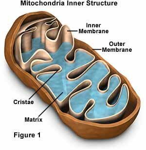
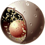
The mitochondria have an outer membrane and an inner membrane that is thrown into folds called cristae. The mitochondria contains the enzymes that carry out the Krebs Cycle, the metabolic pathway involved in the oxidation of the food we eat. These enzymes are in the fluid part of the matrix. Many protein complexes containing cytochromes (which resemble hemoglobin and contain iron) are embedded in the inner membranes. They are responsible for the transport of electrons from the food we eat to the oxygen we breathe. The ultimate site of oxygen utilization is, therefore, in our mitochondria. In the process of shuttling electrons from one cytochrome complex to the next, ATP is produced. We need a lot of ATP just to stay alive and we need even more to engage in the myriad of activities we engage in every minute of the day. You will find mitochondria in all eukaryotic cells but you may find more in those cells which high energy needs such as those in the central nervous system (CNS), muscles, and kidney. The tail of a mammalian sperm is a flagellum made of microtubules around which many mitochondria are wrapped to provide energy for flagellar movement. Mitochondria contain prokaryotic type DNA (single double stranded and circular) and prokaryotic type ribosomes (smaller than the cytoplasmic eukaryotic ribosomes). While dependent on some nuclear gene products, they make many of their own proteins on their own ribosomes from their own DNA messenger RNA and transfer RNAs. However, some of the mitochondrial proteins are coded for by nuclear DNA genes. In humans there are a number of mitochondrial disorders that are due to mutations in either the nuclear genes that provide these proteins or mutations in the mitochondrial DNA itself. Some of these mutations cause deafness, blindness, generalized weakness, diabetes, and many are fatal.
Chloroplasts also have double membrane but instead of having the inner one thrown into folds, there are stacks of disk shaped membranes where the light reactions of photosynthesis occur. The light reactions require the movement of electrons that are activated by light and the proteins that shuttle the electrons are once again embedded in membranes for the smooth flow of the electrons and the production of ATP. The fluid matrix contains the enzymes required for the so-called dark reactions of photosynthesis where glucose is synthesized from carbon dioxide, ATP, and the electrons from the light reactions. Chloroplasts, too, have prokaryotic DNA, prokaryotic ribosomes and their own transfer RNAs. They also make some of their own proteins but the host cell DNA also codes for many of the proteins found in chloroplasts.
Comparison of Prokaryotes and Eukaryotes
| Prokaryotes | Eukaryotes | |
| Organisms | Monera: Eubacteria and Archebacteria | Protists, Fungi, Plants and Animals |
| Level of organization | single celled | single celled (protists mostly) or multicellular usually with tissues and organs |
| Typical cell size | small (1 -10 microns) | large (10 - 100 microns) |
| Cell wall | almost all have cell walls (murein) | fungi and plants (cellulose and chitin); none in animals |
| Organelles | usually none | many different ones with specialized functions |
| Metabolism | anaerobic and aerobic; diverse | mostly aerobic |
| Genetic material | single circular double stranded DNA | complex chromosomes usually in pairs; each with a single double stranded DNA molecule and associated proteins contained in a nucleus |
| Mode of division | binary fission mostly; budding | mitosis and meiosis using a spindle; followed by cytokinesis |