BIO 442 MENU
syllabus 
1 - genome
2 - cell cycle
3 - karyotype
4 - chromab
5 - sexdeterm
6 - prenatal
7 - mutation
8 - mendelian
9 - complex
10 - non-trad
11 - clinical
12 - newborn
13 - teratog
14 - linkage
15 - DNA prof
16 - quanti
17 - links
18 - quizzes
(full title of lecture appears in status bar on the top or at the bottom of your window)

Biology 442 - Human Genetics
The Cell Cycle, Mitosis and Meiosis and Non Disjunction
Cell Cycle
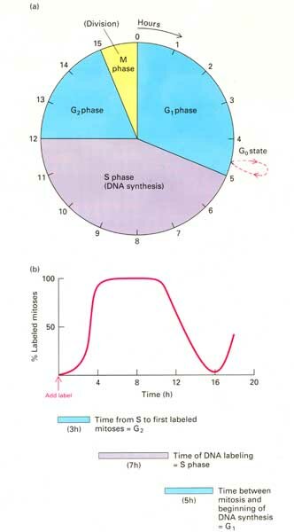
The cell cycle describes the various stages through which a dividing cell passes. After mitosis, a cell goes into G1 (growth 1 phase) during which it increases in size. Most cells in your body are not dividing and remain in a phase called Go. Those cells that divide such as those in the lining of your digestive tract, your skin, the lymphocytes, etc., go through what is known as the cell cycle. They leave Go or G1 and enter the S (synthesis) phase when they replicate their DNA and histones and each chromosome forms chromatids. After the S phase they enter G2 (growth 2 phase) in which they assemble the spindle proteins, etc. in preparation for mitosis or meiosis. When cytogeneticists prepare cells for high resolution banding (HRB) they add methotrexate, a drug which inhibits DNA synthesis, to the cell culture medium. This accumulates cells at the point prior to the beginning of the S phase (sort of like lining horses up at the gate). After an appropriate time (the predetermined length of G1), they add thymidine to the medium to allow the culture cells to enter S phase together and they will stay synchronized for a while. Because they have previously determined the time of each phase (S and G2) they know when the synchronized cells will enter prophase. They then "fix" the cells when the chromosomes are in pro-metaphase and maximally extended. These extended chromosomes when fixed and stained by Giemsa trypsin banding (GTG) will have the maximum number of bands. This procedure allows the cytogeneticist to see some of the larger microdeletions.
Comparison of Mitosis and Meiosis
After fertilization, mitosis gives rise to the embryo and fetus. Most cells in your body are not dividing. When a cell divides it goes through the cell cycle which is guided by cyclins and cyclin dependent kinases. Mutations in genes for any of these or other proteins that control the cell cycle can result in cancer. Incidentally, cancer arises from one or more somatic mutations in one cell. Therefore, cancer is "clonal" in origin as we will discuss later. Meiosis or gametogenesis occurs only in the gonads. It is called spermatogenesis in the male and oögenesis in the female. There are differences between the oogenesis and spermatogenesis which you will learn.
Both mitosis and meiosis require DNA and histone synthesis prior to cell division. Meiosis involves two cell divisions and requires the pairing of homologs in prophase I. This specific pairing is responsible for the orderly segregation of homologs at anaphase I and the recombination of genes between the homologs in pachytene of prophase I. Pairing and crossing over between homologs (at least once cross over per arm) is a requirement for successful completion of meiosis. Even if no crossing over occurred in meiosis and we had to rely on the segregation of homologs at metaphase I of meiosis for random assortment, the probability that two gametes would even contain the same complement of centromeres is 2n where n = 23, the number of chromosome pairs. Stated another way, the number of different gametes due to segregation at Meiosis I would be 223, a very large number! With crossing over, of course the number becomes significantly larger.
To understand the difference between mitosis and meiosis it may be helpful to understand about the infertility of mules and seedless watermelons! Mules are the result of a cross between a horse (2n = 64) mother and donkey (2n = 62) father, hinnies are the results of the opposite cross, donkey mother, horse father. Since the fertilized egg produces the offspring by mitosis the fact that there are not homologous chromosomes in the mule presents no problem. However, when the mule (male or female) attempts to undergo meiosis to produce gametes, the lack of homologous chromosomes becomes a problem since successful meiosis requires the pairing up of homologous chromosomes. No homologues, no meiosis, no baby mules. Seedless watermelons are the results of a cross between a tetraploid (4n) watermelon (gametes are 2n) and a diploid (2n) watermelon (gametes are n) which gives rise to a triploid (3n) watermelon. The triploid watermelon arises by mitosis but when it attempts meiosis, it fails because there are three instead of two homologous chromosomes. Therefore, runty little seeds instead of the characteristic large black ones are produced. In mitosis all chromosomes go to the metaphase dance alone; in meiosis, chromosomes must find a (single) partner with whom to go to the metaphase I dance.
The differences between mitosis and meiosis are clearly seen in prophase. Prophase I of meiosis has several stages LZPDD(D): Leptotene chromosomes are elongated and not yet in pairs; in zygotene the homologous chromosomes form bivalents; in pachytene the synaptonemal complex is formed between the thickened chromosomes and recombination or crossing over occurs; in diplotene, the homologs separate but are held together by chiasmata, one per arm, which are the result of crossing over; in diakinesis, the bivalents are more contracted; and, in females, there is a prolonged stage called dictyotene which lasts until the egg is ovulated. Homologous chromosomes separate in metaphase I and chromatids separate in metaphase II. In females, Meiosis I is completed only at the time of ovulation and Meiosis II only after fertilization. There are several million eggs at the time of birth (held in dictyotene) in the human female. However, the eggs degenerate over time (thank goodness!). There are more crossing over events in oogenesis than spermatogensis. However, there is more recombination in the telomeric regions of chromosomes in male meiosis than in female meiosis. Crossing over is avoided in both near LINE sequences and cytosine methylation sequences. There is also more non disjunction in oogenesis than in spermatogenesis. Non disjunction occurs most frequently at metaphase of Meiosis I and less frequently at metaphase of Meiosis II. Non disjunction is associated more frequently with advancing maternal age.

PACHYTENE
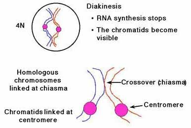
Male meiosis begins at puberty and does not stop. Billions of sperm are produced. Each of the four gametes resulting from each meiotic event is differentiated into a mature sperm. In male pachytene, the X and Y pair at the pseudoautosomal region at the tip of the p arm. Because of the large number of mitotic and meiotic events that occur in spermatogenesis, new mutations are more frequently associated with advancing paternal age.
As mentioned previously, oogenesis begins in the female during fetal life. Meiosis I is completed at ovulation and Meiosis II at the time of fertilization. Only one functional gamete is formed since most of the cytoplasm goes to one cell, the egg. The first polar body contains none of the same information found in the egg; the first polar body does not always go through Meiosis II. The second polar body is more similar genetically to the egg since it receives the other chromatid which differs only where crossing over has occurred. [If a first polar body did go through meiosis II and was fertilized, the resulting twins would be less alike than DZ twins. If the second polar body was fertilized, the resulting twins would be more similar than DZ twins.]
Mosaicism
Non disjunction can occur in mitosis as well as meiosis. When it occurs in mitosis, it is referred to as post zygotic non disjunction and it results in mosaicism. This can occur in the embryo or later. Mosaicism refers to the presence in an individual of genetically different cell lines which arise from a common cell. This can be a new mutation or a chromosome abnormality. It is almost a certainty that each of us have cells with new mutations and/or abnormal chromosomes somewhere in our body. You could have trisomy 21 in your big toe! The consequences of these genetic changes depend on when and where in our development the mutation or non disjunction occurred. Autosomal monosomic cell lines usually die out.
Summary of Differences between Male and Female Meiosis
Human male meiosis (spermatogensis) differs from female meiosis (oögenesis) in several respects:
1. Oogenesis begins meiosis in 3rd month of fetal life; 4th month to zygotene; 7th month to diplotene; 9th month to dictyotene (stage unique to eggs); 2 x 10⁶ oocytes at birth, 90% degenerate by puberty. Ovulation occurs once per month between 12 and 50 years of age. Ovulation stimulates completion of Meiosis I. The first meiotic division produces the first polar body which contains the homologous chromosomes of those in the egg. Preimplantation genetics uses analysis of 1st polar body (2n). If mom is a heterozygous carrier and there are 2 copies of the mutant gene in the 1st polar body, the egg will be okay. If 1 normal copy and 1 mutant copy are found then you do not know if the second polar body (n) or the egg (n) received the mutant copy so you must analyze the second polar body. By "subtraction" one can tell what gene the egg received.
2. Unequal division of cytoplasm in oogenesis between egg and polar bodies.
3. Fertilization triggers the egg to complete Meiosis II and to produce the second polar body.
4. Spermatogenesis produces 4 gametes per meiotic event; oogenesis produces one gamete and 2 or 3 polar bodies
5. There is more crossing over in oogenesis than in spermatogenesis.
6. There is more crossing over near the telomeres in males than in females.
7. There is more non disjunction with increasing age in oogenesis (meiosis I).
8. There are more new mutations with increasing age in spermatogenesis. Advanced paternal age is associated with an increased risk of new mutations. The risk increases linearly with age. The increased risk is related to advanced age of the father for AD conditions and the maternal grandfather for X-linked conditions. Family histories will show no affected individuals since these mutations are sporadic.
New Mutations: Sporadics
Examples of new mutations: AD Osteogenesis Imperfecta Type III and Cornelia de Lange in two newborns, achondroplasia (short limbed dwarfism) in a 2-year-old female with unaffected parents and a case of XR oto-palatal-digital syndrome in a 2 year old male with an unaffected sib and a mother with no family history of affected male children of her mother or sisters. New spontaneous mutations have a small but finite risk of recurrence (0 - 7%) due to possible gonadal mosaicism. The American College of Medical Genetics has issued a Statement on Guidance for Genetic Counseling in Advanced Paternal Age which is available on their Web site: http://www.faseb.org/genetics/acmg/pol-menu.htm
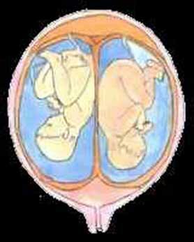
Twins
Monozygotic twins (MZ), are clones and the only examples of natural human genetic identity (besides clones). Dizygotic (DZ) twins are no more similar genetically than sibs since they come from two different eggs and two different sperm. DZ twins result from multiple ovulations and have been known to have two different fathers. If inherited, it is most commonly inherited through the maternal lineage due to multiple ovulation. Multiple fetuses including twin pregnancies are usually considered high risk because of fetal crowding and commonly premature delivery.