BIO 102 MENU
syllabus 
1 - origin
2 - biomol.
3 - biomol2
4 - viruses
5 - prokaryon
6 - endosym
7 - eukaryon
8 - energy
9 - mitosis
10 - meiosis
11 - reprod
12 - genetics
13 - humgene
14 - humge2
15 - evolution
16 - evolutio2
17 - diversity
18 - diversi2
19 - tissues
20 -digestive
21 - respirat
22 - circul
23 - excret
24 - endocr
25 - receptors
26 - nervsys
Quizzes
Bio 103 Lab
(full title of lecture appears in status bar on the top or at the bottom of your window)

Biology 102 - General Biology
Human Reproduction and Development
Reproduction
All living things reproduce....viruses, bacteria, amoebae, plants, animals, etc. Sometimes the new individual is an exact copy of the parent, except for new mutations, and sometimes it is the product of two parents. We have two parents but a bacterium or amoeba has one parent who disappears when it is formed! Exact copies are formed by mitosis (asexually) in eukaryotic organisms. Prokaryotes need only replicate their single DNA molecule to make new organisms like themselves. All organisms including viruses, Monera (prokaryotes), Fungi, Protista, Plants and Animals have sexual reproduction capability whereby genetic information from more than one parent contributes to the new organism. Plants, for example, can send out runners or you can stick a stem in the ground and a new plant will form by mitosis from the "parent" plant. Humans (and other mammals) only reproduce sexually but if cloning becomes common then we will have asexually produced offspring! Sexual reproduction assures that the offspring are different from the parent(s).
Most animals and plants have complex organs to assist in sexual reproduction. Sometimes these are in different individuals, male and female. Sometimes one individual has both male and female gonads. Gonads are specialized organs called testes and ovaries in mammals and some other organisms. Meiosis occurs in the gonads to make gametes (sex cells) called sperm and eggs. Plants make pollen in the stamens (male parts) and ovules in the ovary (female part). There are many variations on this basic theme of making male and female gametes. The formation of male and female sex organs and gametes is under genetic and/or environmental control in organisms. While we will focus on sexual reproduction and development in humans, much of what you will learn applies in general to mammals in general.
In humans, sex can be defined on several levels: chromosomal sex (XX or XY); gonadal sex (testes or ovary); internal plumbing (oviduct, uterus, cervix, vagina or epididymis, vas deferens, urethra) and external plumbing (labia minora, labia majora, clitoris, vagina or penis, scrotum); and psycho-social sex (sometimes referred to as gender). For the detailed notes on sexual differentiation, go to the Bio 442 lecture note on my web site (carolguze.com) Lecture on Sex Determination.
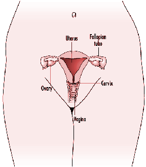
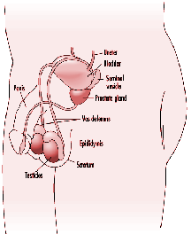
HUMAN REPRODUCTIVE SYSTEMS: GONADS (OVARIES AND TESTES) AND DUCTS
Development
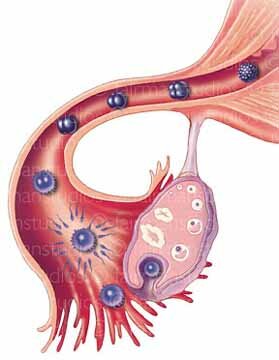
Eggs are formed and meiosis begins in the human female fetus early in development but the egg is arrested in Meiosis I until it is ovulated. Once a month, an egg is released from a ripened follicle beginning at puberty and continuing until menopause which occurs when a female is in her 40's or 50's. These processes are under hormonal control. Meiosis and sperm production in the male, unlike in the female, does not begin in the male until puberty but continues throughout his lifetime. Fertilization is the union of a sperm and egg. This occurs high up in the oviduct in humans. When the egg is released from the ovary in humans, it completes Meiosis I. Meiosis II is completed when the sperm penetrates the egg. After fertilization, when the egg is still in the oviduct, it begins to divide by mitosis to form a hollow fluid filled ball of cells called the blastula. This process is called cleavage. When it reaches the uterus (womb) 6 or 7 days after fertilization, it embeds (implants) in the uterine lining (endometrium). Sometimes a little blood is released as it burrows into the endometrium and some women mistake this blood for a menstrual period and, therefore, are off by a month in determining when they became pregnant. To maintain the endometrium, the blastocyst cells secrete the hormone hCG (human chorionic gonadotropin). This can be detected in the mother's blood or urine at about 3 weeks and it is hCG that is detected in home pregnancy test.
The embryo proper begins to form from an inner cell mass within the blastocyst. Some parts of the blastocyst will form the extra embryonic membranes such as the amnion, chorion and parts of the placenta. The embryo forms on a plate-like layer of cells. It begins with a "primitive streak" which establishes the anterior and posterior axes and the bilateral symmetry of the body. The neural tube forms from the union of two ridges on the top side. As is true for all vertebrates, 3 cell layers are formed these layers are called the ectoderm, mesoderm and endoderm. These form by cell divisions, migrations, and rearrangements and are the forerunners of specialized tissues and organs. The ectoderm is the outermost primary tissue layer that forms first in all animal embryos. The ectoderm will give rise to the tissues of the nervous system and the structures that form the outer surface such as hair and teeth. The endoderm is the innermost layer, it forms the gut and structures derived from the gut. The mesoderm is the intermediate layer (last to arise evolutionarily) and it is important in the formation of almost all organ systems including the muscles, skeleton, and connective tissues. After these layers form, subpopulations of cells give rise to the organs. The final stage of development is growth and tissue specialization.
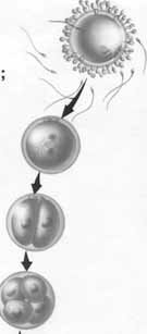

AFTER FERTILIZATION IN THE OVIDUCT, THE EGG BEGINS TO DIVIDE BY MITOSIS. HERE WE SEE THE FOUR CELL BLASTULA. EVENTUALLY THIS ONE CELL WILL GIVE RISE TO THE TRILLION CELLS IN OUR BODY

The formation in the gastrula of the 3 major layers: ectoderm, mesoderm and endoderm


Teratogens are environmental agents that cause developmental defects in the fetus. Examples of teratogens are alcohol, drugs, seizure medications, rubella virus, and accutane. The sensitivity to teratogens is graphically represented in the chart above. The word, teratogen, is derived from "terato" meaning monster and "gen, " to give rise to, so teratogens give rise to monsters (not really). Teratogens are non-genetic factors that interfere with normal embryonic and fetal differentiation and morphogenesis. They are not mutagens. Mutagens act randomly on all DNA and do not produce one specific genotype. Children who have been exposed to teratogens in utero will not pass their defect on to their children. Because the effects of teratogens are seen at birth and are therefore a congenital defect, they are often thought to be genetic and can mimic genetic disorders. But you need to know that congenital refers to showing up at birth and does not mean genetic and genetic does not mean congenital.