BIO 102 MENU
syllabus 
1 - origin
2 - biomol.
3 - biomol2
4 - viruses
5 - prokaryon
6 - endosym
7 - eukaryon
8 - energy
9 - mitosis
10 - meiosis
11 - reprod
12 - genetics
13 - humgene
14 - humge2
15 - evolution
16 - evolutio2
17 - diversity
18 - diversi2
19 - tissues
20 -digestive
21 - respirat
22 - circul
23 - excret
24 - endocr
25 - receptors
26 - nervsys
Quizzes
Bio 103 Lab
(full title of lecture appears in status bar on the top or at the bottom of your window)

Biology 102 - General Biology
Animal Structure and Function
The Circulatory System

The circulatory system usually works in close association with the respiratory system except, as previously noted, in insects. The circulatory system has several functions but primary is the function of transport: transport of gases, nutrients, hormones, toxic, and excess molecules.
It also functions as an internal support (skeleton) in some organisms (e.g., worms) with no endo or exoskeleton but also in humans where it serves to produce the erection in the penis (however, some mammals have an os penis). The circulatory system in some vertebrates has protective functions. It contains the white blood cells which fight infection and it contains cells which release proteins which cause clotting to protect from "leaks."
HOW WE FIGHT INFECTIONS WITH ANTIBODIES MADE BY THE IMMUNE SYSTEM
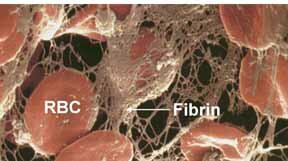
BLOOD CLOT
The circulatory system consists of a heart (or hearts) which is a bigger, pumping muscle and blood vessels. Some vessels lead away from the heart (arteries) and some toward the heart (veins). The arteries have thicker walls than the veins. The veins can be distinguished from arteries by the fact that they have valves to prevent back flow of the blood. The arteries and veins branch and become narrower until they meet in a capillary bed. The capillaries are where the action is! These vessels are only one cell thick and therefore gases, nutrients, hormones, toxic molecules, water, etc. can freely flow in and out. Also white blood cells can squeeze out between the cells lining the capillaries (endothelial cells). Red blood cells carry oxygen and release it in areas of low concentration. Conversely, they pick up carbon dioxide from areas of higher concentration and carry it to be released in areas of lower concentration. Gas exchange occurs simply by diffusion from areas of higher concentration to those of lower concentration no ATP is used!
The insects, who have no need for an efficient circulatory system, have what is called an open circulatory system. They have a heart which pumps the blood into open-ended arteries and the "blood" sloshes around to reach the cells of the body. It is passively recollected by open-ended veins to be returned to the heart. We have such an open-ended circulatory system in our lymphatic system. It works in parallel with our closed circulatory system to collect excess fluid that remains in the tissues. If blocked, fluid accumulates, especially in the lower limbs and scrotum.
All animals, other than the insects, have a very efficient closed circulatory system. There is a heart which pumps blood to the arteries, arterioles, capillaries, and venules and veins which collect the blood and bring it back to the heart. The heart in fish is a simple tubular, two-chambered organ. In the vertebrate embryo this is how all hearts begin their development.
Summary of the functions of the circulatory system
- Transport
- H2 O and nutrients from the intestine to the cells or to a storage site.
- O2 from the respiratory organ to the cells and CO2 from the cells back to the respiratory organ.
- hormones from endocrine glands.
- toxic or waste molecules to the excretory organ.
- Protection
- of the organism from foreign invaders (immune system)
- of itself from loss of blood (clotting mechanism)
- Skeletal, especially in some invertebrates.
Respiratory Proteins Carry Oxygen to the Body Cells
Since O2 is not readily soluble in water, animals whose circulatory system works closely with the respiratory organs require special respiratory proteins in their blood...either free in the blood or in blood cells. We carry our respiratory protein, hemoglobin, in our red blood cells (rbcs). The protein must bind reversibly to O2. (The reason carbon monoxide (CO) is lethal is that it binds irreversibly to hemoglobin.)
Different organisms have different respiratory proteins that work in conjunction with a metal ion. There are two main types. Some use copper (Cu, e.g., hemocyanin) and others use iron (Fe++, e.g., hemoglobin). The iron ion in our hemoglobin is held in a porphyrin group called heme. The protein part of the molecule varies between species and even between the fetus (HbF) and the adult (HbA). Fetal hemoglobin is coded for by a different gene but is very similar in amino acid sequence to adult hemoglobin. It arose by gene duplication and since it bestows an advantage to the fetus, it was selected for during the evolution of the mammals. The adaptive value of HbF is that it binds oxygen more tightly than HbA and is, therefore, able to "suck" the oxygen from the mother's blood stream.
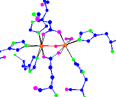
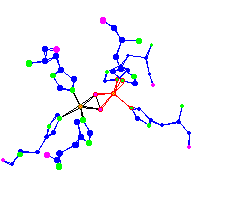
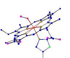
Hemerythrin Hemocyanin Hemoglobin
BLOOD PROTEINS FOUND IN INVERTEBRATES (hemerythrin and hemocyanin) AND VERTEBRATES (hemoglobin)
Proteins that combine with O2 are also found inside cells. The muscle cells have the heme containing protein, myoglobin, which, like HbF is able to draw the O2 away from the hemoglobin in the red blood cells. The ultimate destination of O2 is cytochrome C, another heme containing protein, in the electron transport system in the mitochondria.
Blood. Blood is essentially a salt solution similar to the intracellular fluid and osmotically balanced with the intracellular fluid. In addition to the inorganic ions (K+, Na+, HCO3-, HPO4-, Cl-, Mg2+, Ca2+, etc.) there are protein molecules. In vertebrates the blood contains specialized proteins and cells. Some of the proteins carry molecules such as lipids (HDL and LDL) and ions (transferrin, ferritin). Other proteins include antibodies (gamma globulin fraction) and serum albumin. The specialized cells in our blood are the red blood cells (rbcs) containing the respiratory protein, hemoglobin, to carry gases, the white blood cells (e.g., phagocytes, lymphocytes) which fight infection and the blood platelets which upon disruption release enzymes to catalyze the formation of clots when a "leak in the pipes" is threatened.
The structure of circulatory systems
In simple...small or inactive animals such as sponges, Cnidaria and flatworms....the transport of O2 and CO2, nutrients and wastes is solved simply. The two or three cell layers that form the organism are either close to the external fluid media or the internal fluid of the gut. The rudimentary muscles of the body wall assist in the movement of the molecules needed.
However, in more complex animals with organs and tissues well removed from the sources of gases and nutrients, more is required. Pipes are necessary to convey the molecules to every cell in the organism's body. The organs of circulatory systems are the heart (pump) and blood vessels. The blood is the (connective) tissue that carries the gases, ions and organic molecules. The arteries carry blood away from the heart and the veins carry the blood back to the heart.
(Even the plants that are tall require "pipelines" provided by their vascular tissues. The xylem and phloem are vascular tissues found in complex plants. Their function is to transport water, mineral ions and organic nutrients from roots to leaves and leaves to roots. The plants, however, lack a pump or heart to assist in transporting these molecules.)
Hearts
Hearts are pumps. They are highly muscularized portions of the circulatory system, often with a thin walled atrium, thick walled ventricle and valves to prevent back flow. In animals with one heart, a choice has to be made whether to place it before or after the respiratory organ. If the heart is placed after the respiratory organ (gills) as it is in most invertebrates (e.g., clams lobsters), the blood pressure in the respiratory organ is sacrificed to have a higher blood pressure to the body (systemic circulation). Conversely, if the heart is placed before the respiratory organ as it is in the fish, the higher blood pressure in the gills provides for more efficient gas exchange at the sacrifice of the systemic blood pressure. In the other vertebrates, the oxygenated blood flows back to the heart to be pumped to the entire body (systemic circulation). Since gas exchange is a critical function of a circulatory system, the vertebrates (except the fish) have the heart located before the respiratory organs for maximum flow rate. In cephalopod mollusks (e.g., squid and octopus) there are two gill hearts that pump blood to the gills. The oxygenated blood from the gills flows into another single (systemic) heart which pumps the oxygenated blood to the animal's body.
In mammals and birds (and maybe some reptiles) the heart is actually two hearts side-by-side. The right side of our heart pumps blood to the lungs and the left side collects the blood from the heart and pumps the oxygenated blood to the entire body (systemic circulation). In this way the blood pressure is high for both the pulmonary and systemic circulation.
Our hearts have a thin walled right atrium which collects deoxygenated blood from the body and sends it to the lungs via the pulmonary arteries (with deoxygenated blood). The blood then flows through the capillaries surrounding the alveoli in the lung where CO2 is released and O2 is picked up. The capillaries reform to give rise to the pulmonary vein (with oxygenated blood) which leads back to the left atrium. The blood then flows to the thicker walled left ventricle which pumps the blood under pressure to the body. The freshly oxygenated blood is sent first to the coronary arteries that supply the heart and to the aorta which branches into many arteries which in turn supply blood to all the organs and tissues. The carotid artery to the brain also branches off the aorta near its origin.
When a person has a "coronary" we mean s/he has a block (usually an atherosclerotic plaque) in the coronary arteries that supply the heart with fresh oxygen. When this happens, some of the heart tissue is deprived of oxygen and dies (an MI or myocardial infarction).
In the fetus, the blood comes from the umbilical vein (oxygenated blood from the placenta) to the right atrium. Since the lungs are not functioning yet, they are collapsed and only a small portion of the blood goes to nourish them. In the fetus, most of the blood passes directly from the right atrium to the left atrium through a hole called the foramen ovale, which closes up in time. The blood from the left atrium flows to the left ventricle to be pumped to the fetal systemic circulation. If the foramen ovale remains open after birth, a "blue baby" can result because blood from the left atrium mixes with the blood from the right atrium and overloads the right heart.
In simpler animals there is no heart. Either they use the general body wall muscles (sponges, Cnidaria, flatworms) or muscles of the blood vessels (annelids) to move fluids around the body. These peristalsis-like movements cause the blood to flow mostly in one direction...the direction of the wave...but there is back flow unless there are valves.
Blood Vessels
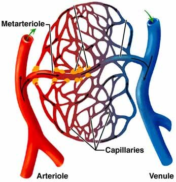
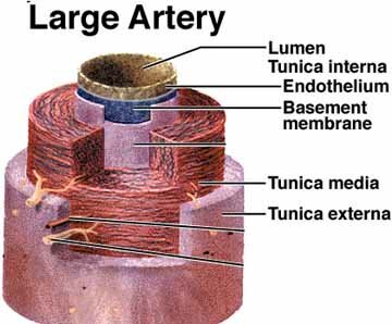
Arteries lead away from the heart and, except in the case of the pulmonary circulation, carry oxygenated blood. They are thick walled with elastic fibers and layers of muscle. Their structure explains how they maintain a high blood pressure. The innermost lining of cells is a one layer thick epithelial tissue called the endothelium. The endothelium continues into the arterioles and finally forms the capillaries. Sphincter muscles at the arteriole-capillary junctions regulate the amount of blood to any organ or tissue. These muscles are controlled by hormones and the nervous system.
The capillaries which are fed by arteries and drained by veins, penetrate to within one or a few cells of every cell in the body. Due to the high blood pressure from the heart and maintained by the arteries, the fluid of the blood is forced out of the capillaries. Everything in the blood except the larger protein molecules and the rbcs can get through the capillary wall, even the amoeboid white blood cells. It is in the capillary beds that the most important events occur. Nutrients and hormones are delivered, wastes picked up, and gases exchanged. As the blood pressure drops, the osmotic pressure increases and the fluid reenters the capillaries.
There is, however, a net loss of fluid and white blood cells in the capillary beds. The lymphatic system, an open circulatory system, supplements our closed circulatory system. The lymph vessels begin in capillary beds and collect into larger vessels that eventually drain into the heart. This system collects excess fluid and proteins (e.g., lymph which is blood minus the rbcs) leaked from the blood into the tissues. It also collects fats from the intestine. Another important role is to bring foreign cells, viruses and other material to the lymph nodes. Lymph nodes are scattered throughout the body. They contain large numbers of lymphocytes, produced in the bone marrow, which fight infections.

THE LYMPHATIC SYSTEM RETURNS EXCESS FLUID TO THE HEART
(It is an "open" system composed of blind ended capillaries that bring lymph to lymphatic veins which return the fluid to the right auricle of the heart. There are lymph nodes along the way and it is part of the immune system.)
Venules are the blood vessels that exit the capillary beds and go on to form veins. The veins are thin walled with a larger bore and with less muscle and elastic fibers than arteries. The flow is slow through them and to prevent back flow, there are valves. The body muscles help keep the blood flowing through the veins. The veins are the final vessels in a closed circulatory system.
In a closed system, as found in the vertebrates and annelids, the blood flows through a continuum of blood vessels. However, in many invertebrates such as the mollusks and arthropods, one finds open circulatory systems. Blood flows through vessels for only part of its path and in part of its path it flows into or out of large sinuses which may bathe the specific organ it reaches. These sinuses serve the same purpose as capillaries and blood may recollect into veins or just return in a haphazard way to the heart. The insects can get by with an open circulatory system because they have the highly efficient tracheal respiratory system that delivers oxygen to their cells.

INSECTS CAN GET BY WITH AN OPEN CIRCULATORY SYSTEM BECAUSE THEY HAVE A COMPLEX RESPIRATORY SYSTEM (TRACHEAL SYSTEM) THAT DELIVERS OXYGEN TO ALL THE CELLS OF THEIR BODY